Pathology
Pathology
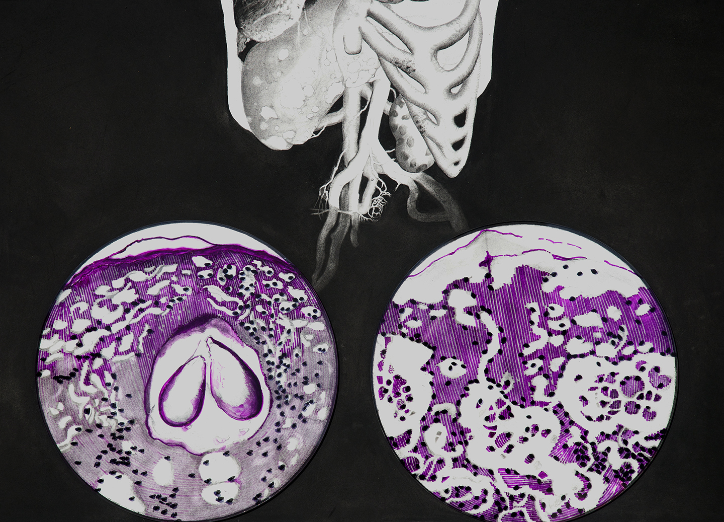
Visualizing Melanoma
Graphite and charcoal on paper with ink on Plexiglas and aluminum mounts
50” x 76” x 4”
Graphite and charcoal on paper with ink on Plexiglas and aluminum mounts
50” x 76” x 4”

From Visual Pathology, a research and exhibition collaboration with the Galveston Art Center and the Department of Pathology at the University of Texas Medical Branch.
My early research at the Truman G. Blocker, Jr. History of Medicine Collections tethered me to Johannis Remmelin’s anatomical ‘flap’ book, Catoptrum Microcosmicum (1667). This is a rare book of full-plate illustrations with layers of paper detailing muscular, skeletal systems, etc. so that lifting the layers shows the anatomy, as it would appear during dissection. Though cutting-edge at the time, I was amused by the hills of wildflowers models stood upon and literal paper fig leaves layered over genitals. I was also looking at Japanese anatomical watercolor paintings from the Edo period and the cross-section illustrations of late-pregnancy by obstetrician William Hunter and engraver Jan van Riemsdyk (Gravid Uterus, 1774).
Expressing interest in cancer’s metastasis in bone and other organs, I received assistance from Dr. Paula Summerly and Mark Demming locating several specimen of malignant Melanoma metastasis in the UTMB Pathology Specimens collection. Each stemmed from a singular case of the foot, including infected clavicles, hands, femur, spleen, and testicles. Though I was immediately compelled to draw these organs, I also needed to learn more about this disease in order to handle the specimen with a high level of educated respect. I began illustrating a full-size torso that would house some of the affected organs and allow for break-off magnified views of affected bone, views of skin cancer in the epidermis and microscope views of Melanoma variations.
While I drew, I strove for anatomical accuracy while realizing that I have a limited familiarity of anatomical organs, especially those affected by disease. My sources ran from the UTMB Pathology collection to Gunther von Hagens’ plastinized Body Worlds exhibit, from carved ivory anatomical “manikins” circa 1500-1700 to medical surgeries live-streaming online. As I learned more about Melanoma it became important to highlight histologic criteria for diagnosing melanoma from other skin carcinoma (basal cell and squamous cell carcinomas), and to show the visual variations that can be identified with a microscope between acral lentiginous melanoma, lentigo malgna melanoma, mucosal lentiginous, and superficial spreading types. Making a condensed drawing in a short period of time required a lot of editing, and as with most of my work, I often had to stop myself from going into obsessive detail on any one organ or system.
Having had the opportunity to research with medical experts and artists was the most stimulating part of the Visual Pathology Project. Dr. Tais Saito answered my questions about Melanoma and Histology (study of the skin), and Dr. Summerly, Dr. Afrouzian, and Dr. Walker provided further support and answers. I look forward to future partnerships within the medical world as technology and research strategies become increasingly collaborative, diversified, and creative.
 Installation shot by Roxann Grover