Phantoms of the Great Dying
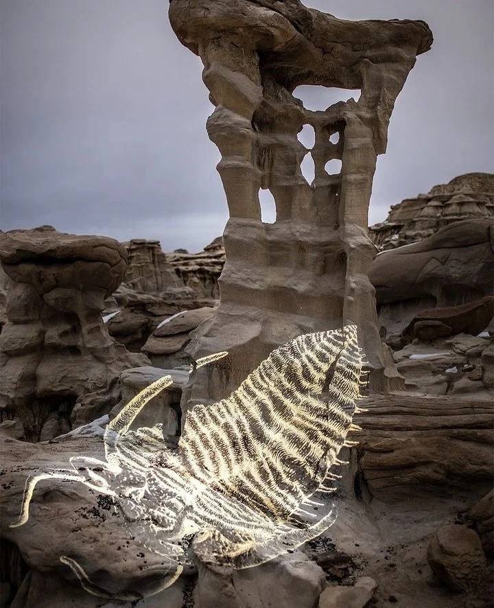
1. Trilobotes Descending the Alien Throne
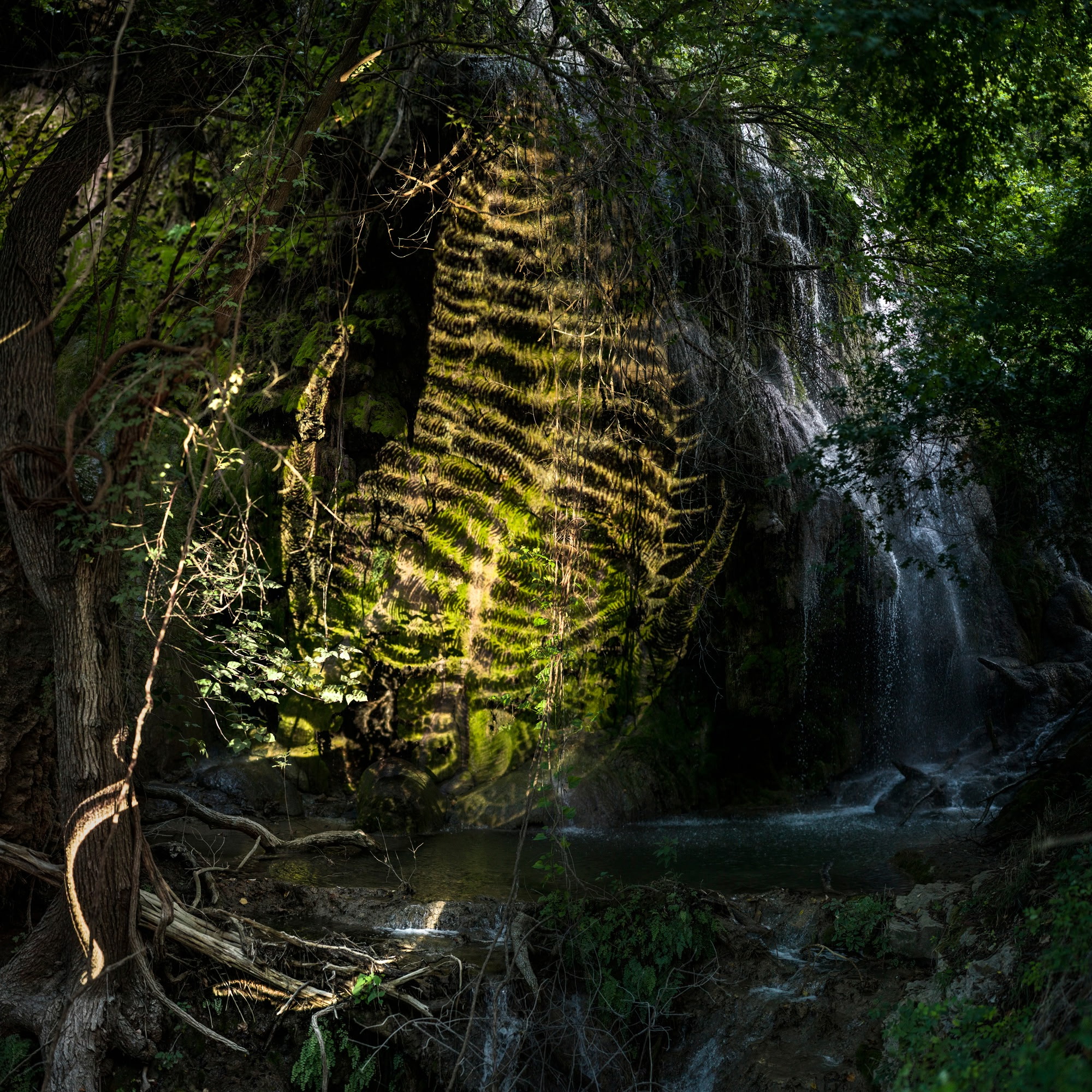
2. Trilobite in the Gorman Falls
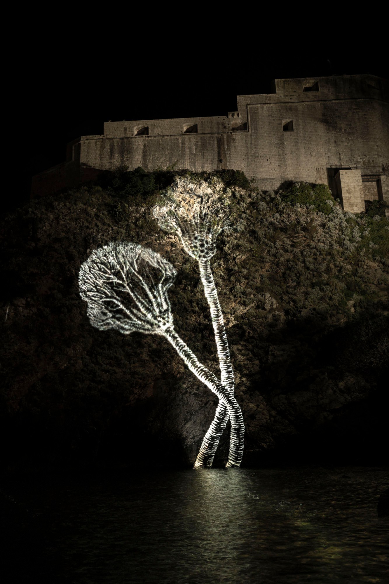
3. Crinoids Reaching for Trdava Lovrijenac
4. Eurypterid at the Bayou
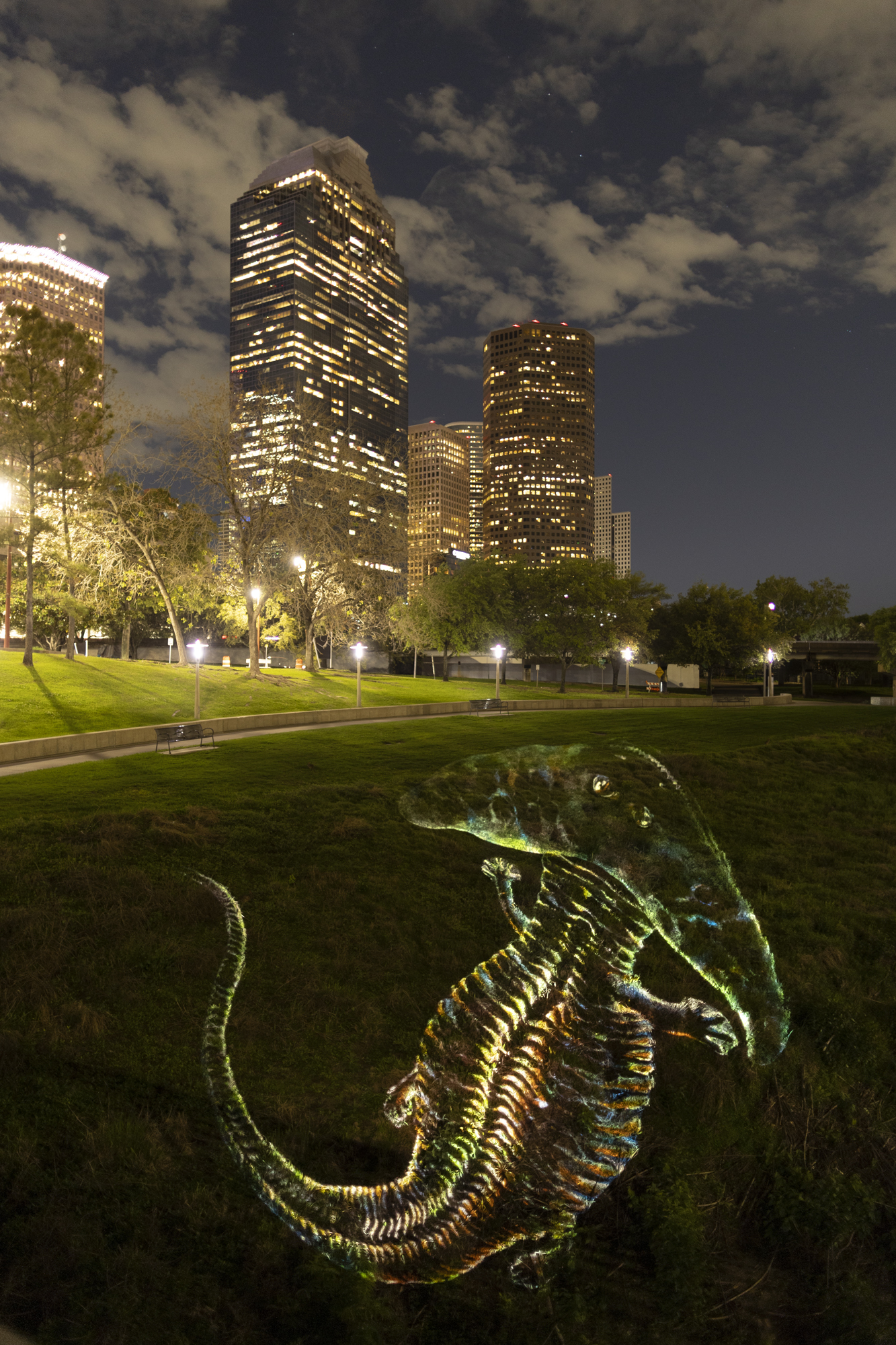
5. Diplocaulus views the Houston skyline
6. Crinoid tours the Arch
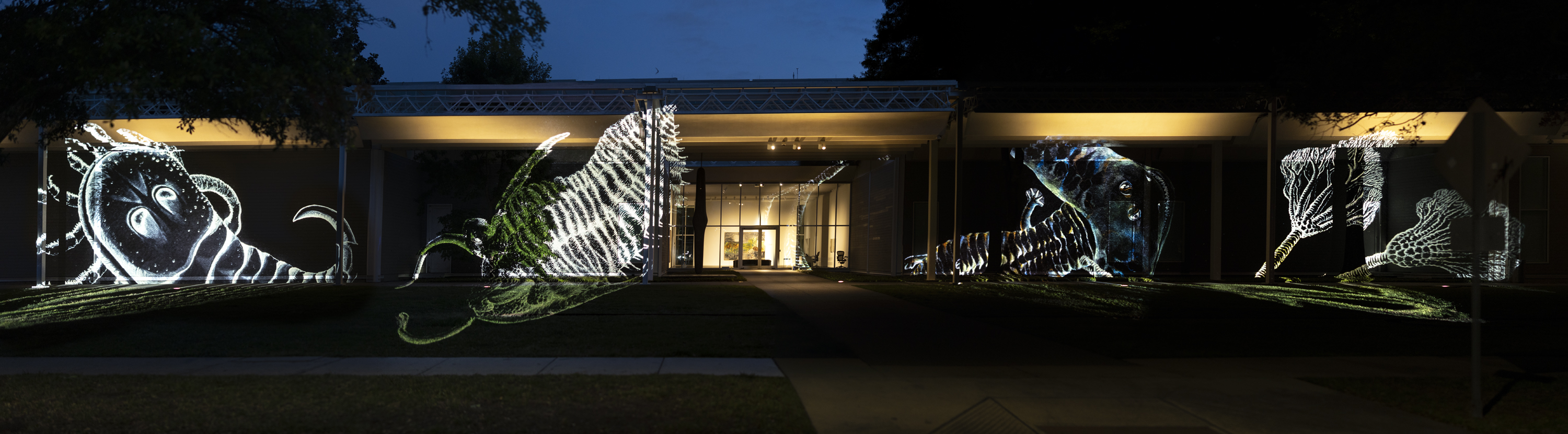
7. Fossils tour the Menil
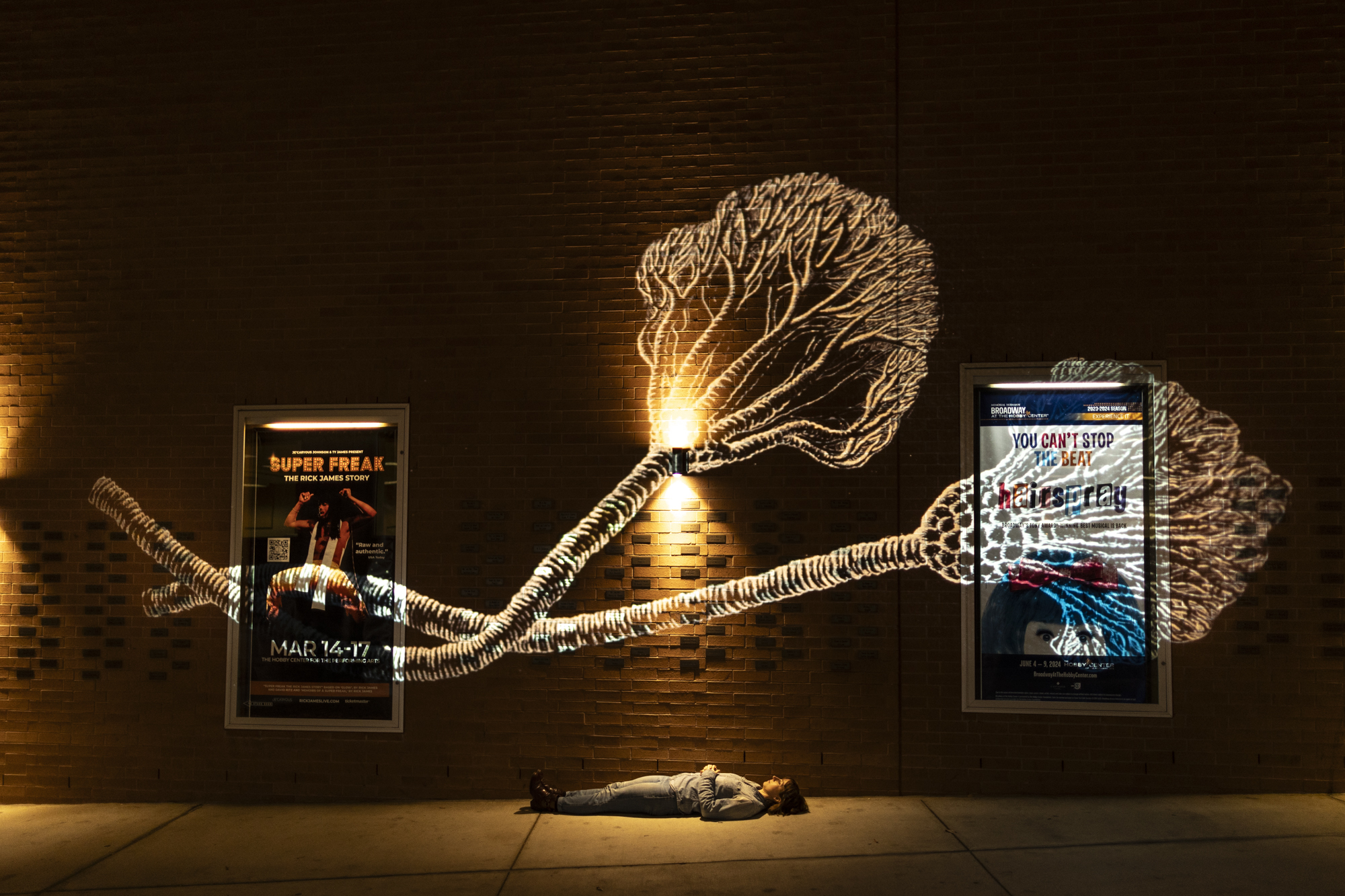
8. Crinoid and the Artist at Hobby Center
9. Drawing process
All images are inket photographs, dimensions variable.
Artists Mark Chen and Colleen Maynard found overlapping interest in deep geological time and evolutionary layers, particularly the Permian-Triassic extinction of 251 million years ago that wiped out nearly 90% of living species at the time. A giant spectrum of plants, fish, amphibians, proto-mammals, arthropods and insects died; among them, trilobites remains became some of the most abundant fossils found globally today. Many other species took such significant hits that they were dethroned from thousands of years in the making. Colleen’s vivid drawings honor the extinct creatures into realized complexity. Mark takes calculated jaunts with his handmade projector to photograph the drawings upon surfaces ranging from mountain bluffs to architectural landmarks.


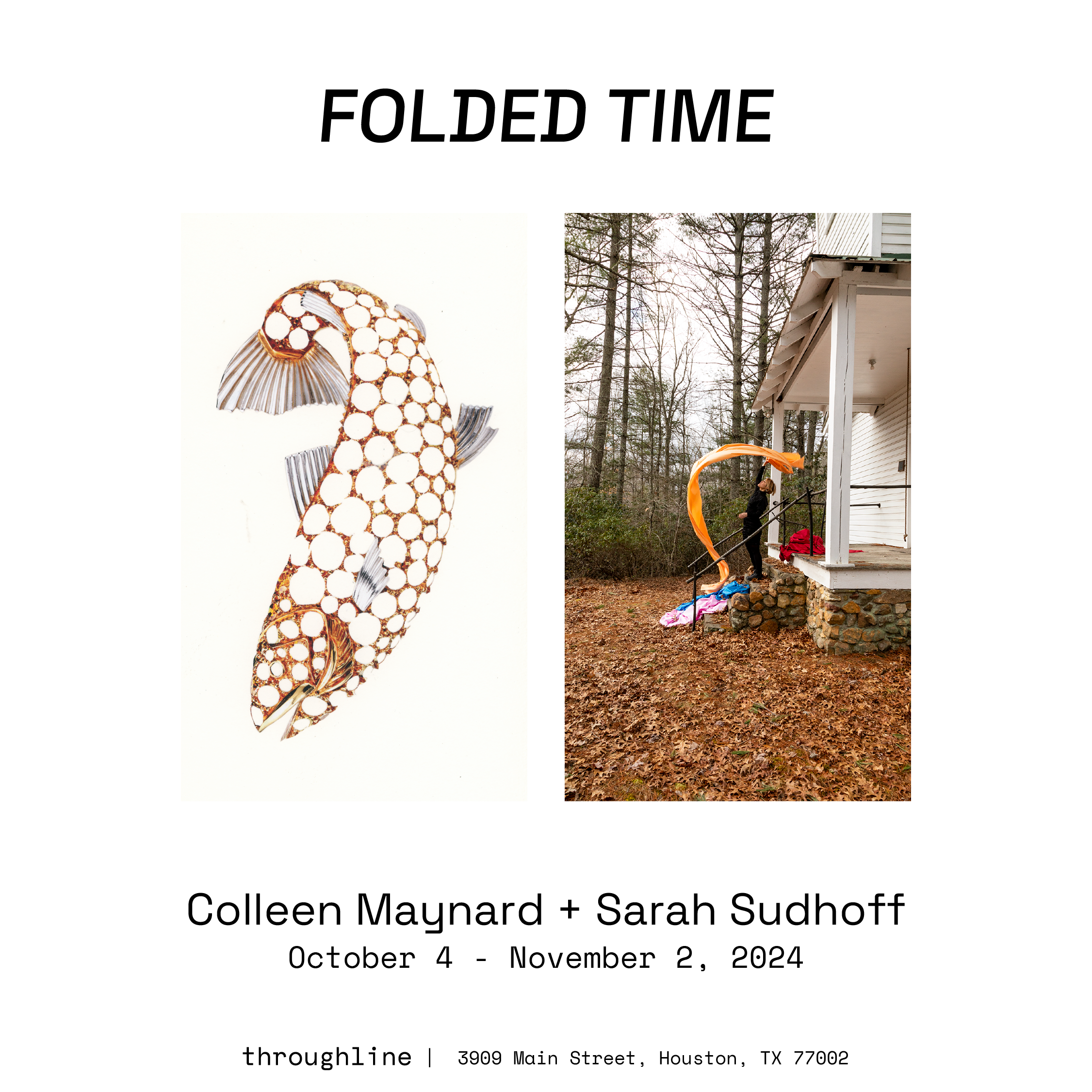
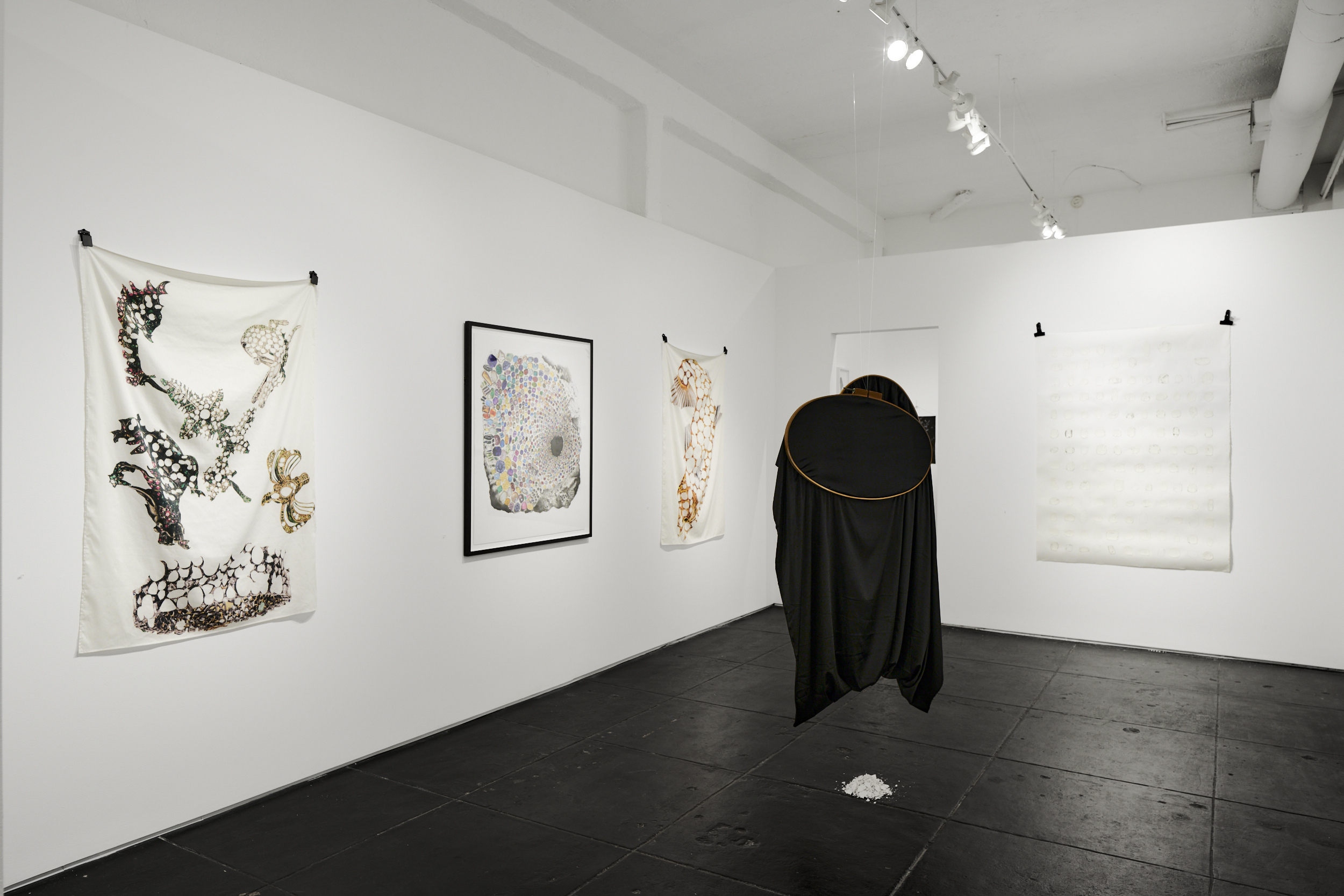
In Folded Time, Colleen Maynard and Sarah Sudhoff examine the compounding and unspooling of deep time and process via performance, drawing, photography and collage. Their interests overlap in both specific physical processes — rituals and customs surrounding death and unique innovations of organisms — and philosophical queries into ways biology, extinction, human mortality and traditions interweave and impact each other.
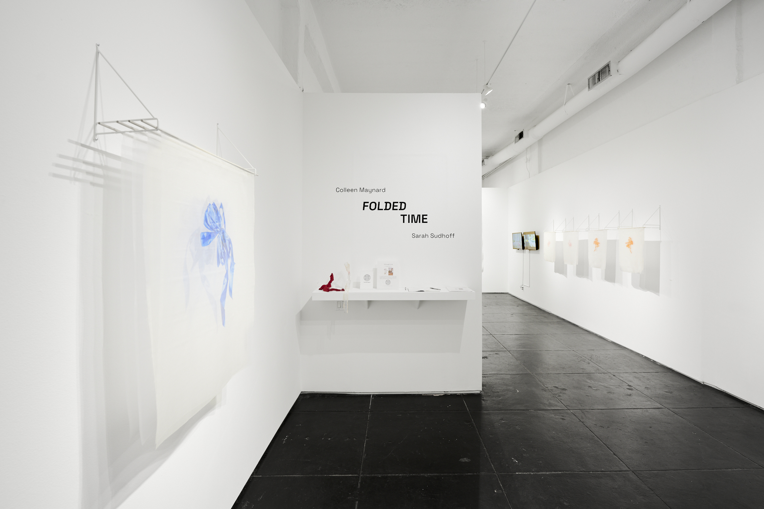
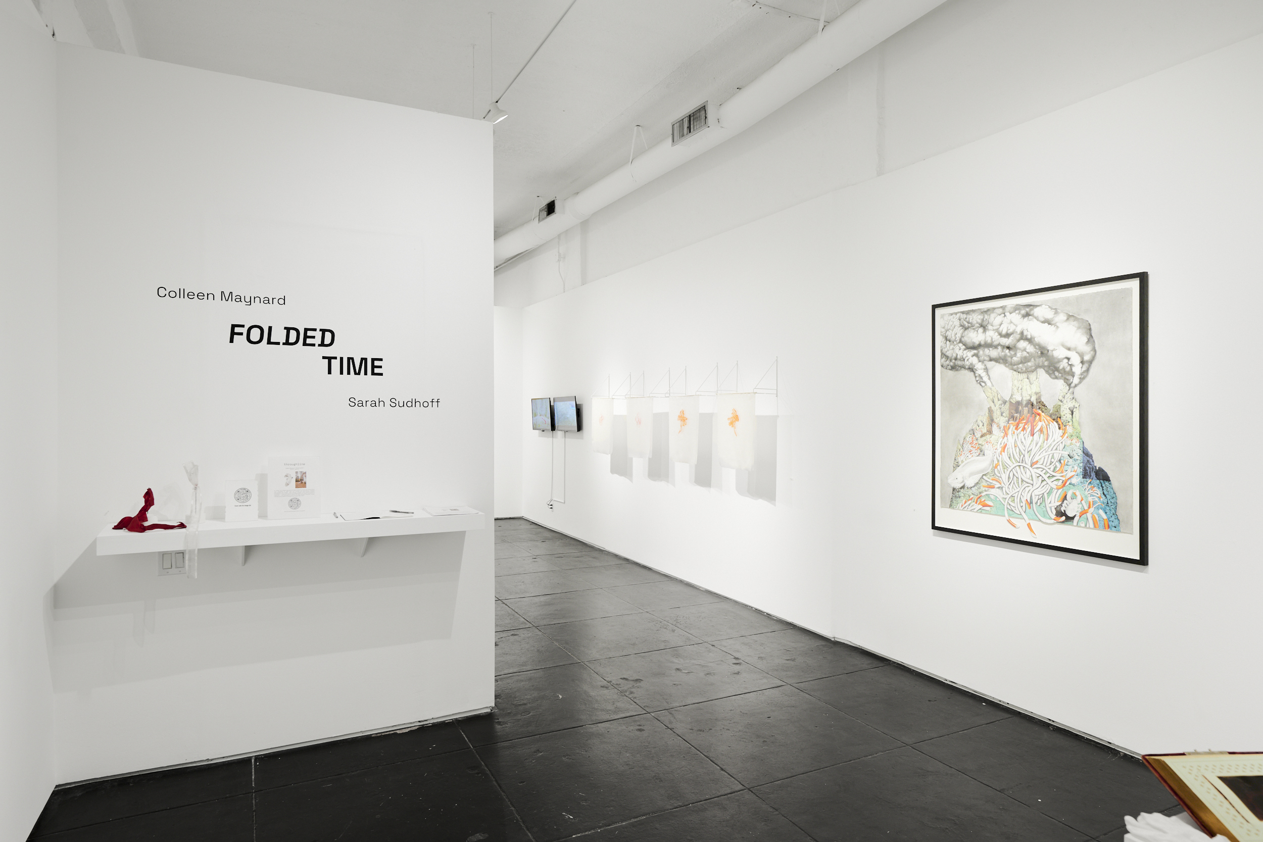
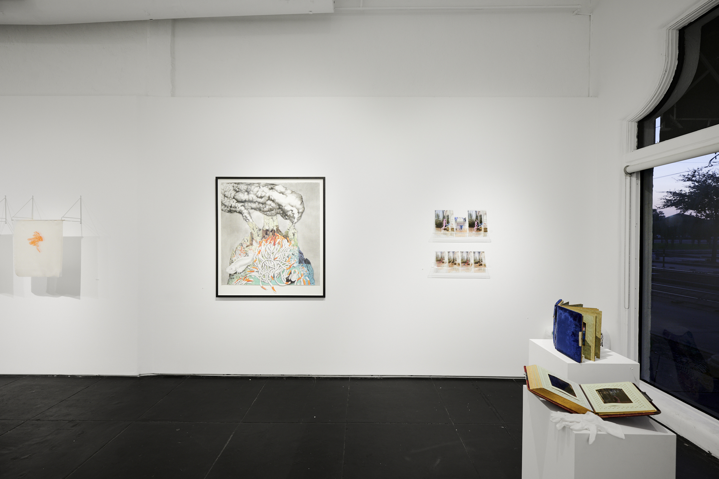
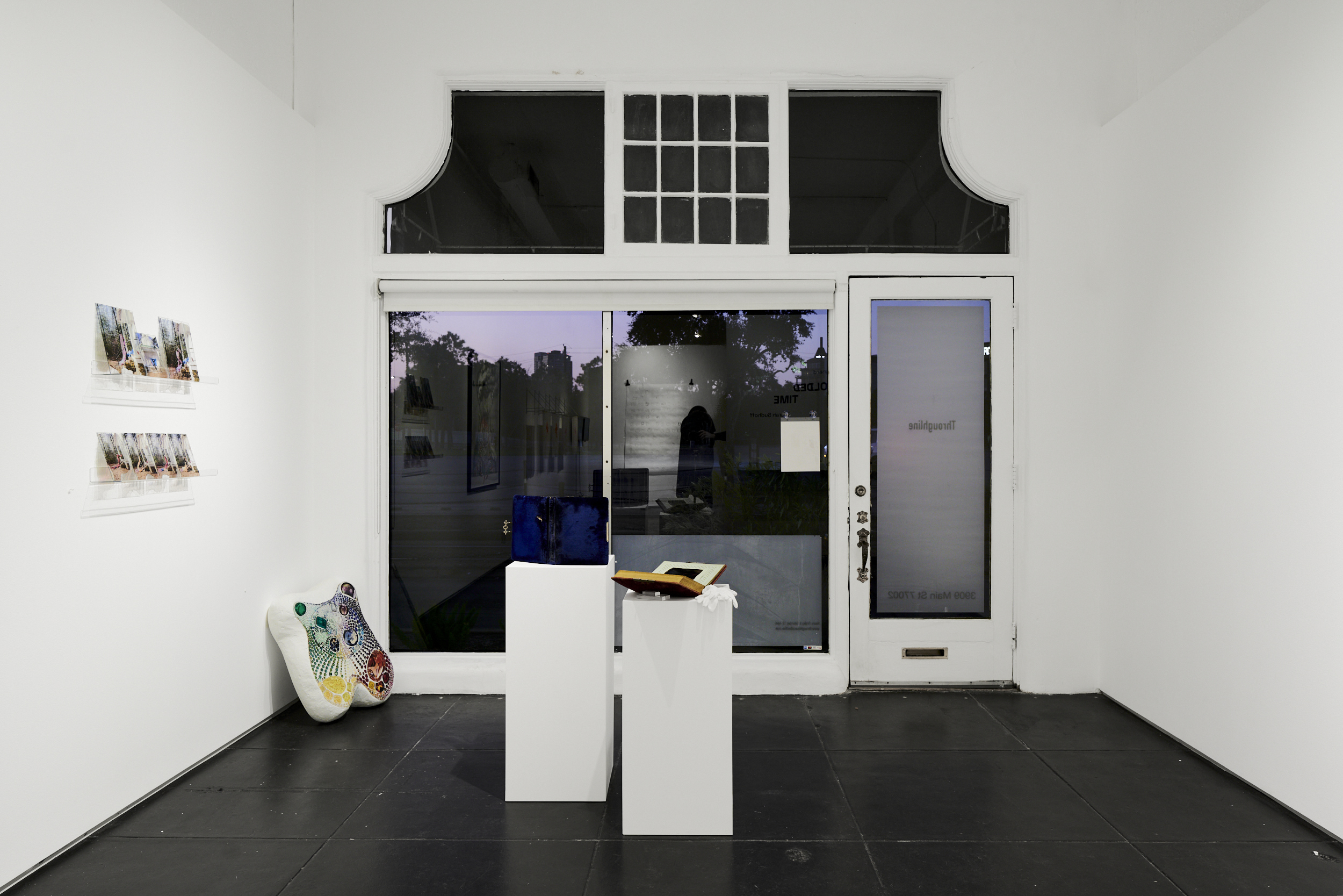
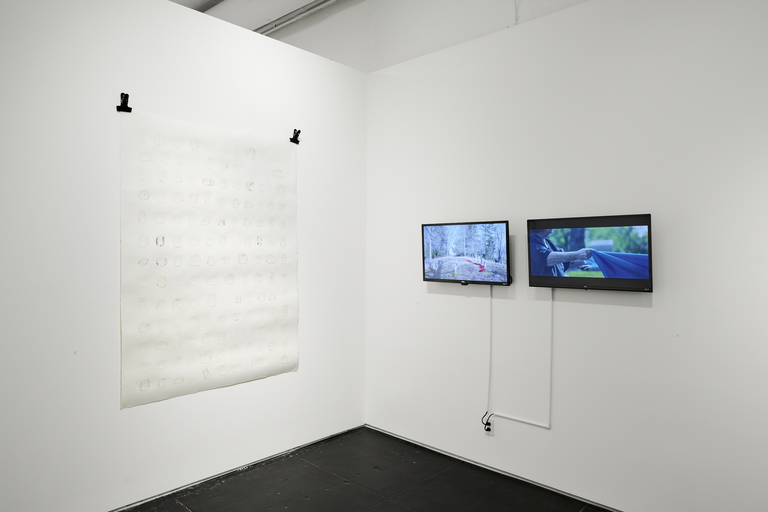


Announcement by Molly Koehn. Video by Beatriz Bellorín. Installation photography by Jake Eshelman (images 1-6).
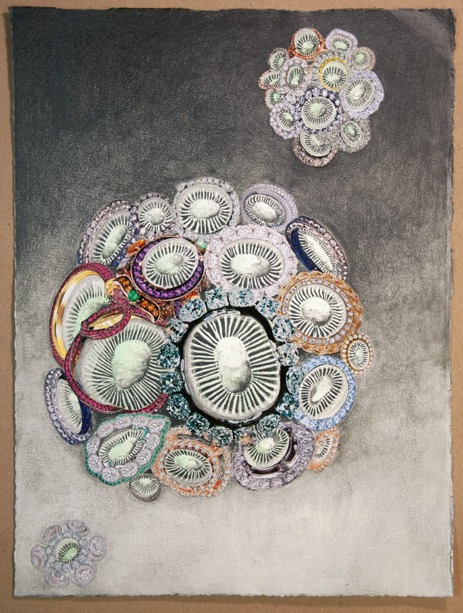
Microcosms
- Coccolithophore (Emiliania huxleyi)
- Lace lichen (Ramalina menziesii)
- Maritime sunburst lichen (Xanthoria parietina)
- Slime mold (Physarum polycephalum)
In middle school I walked my yellow Schwinn bike through a forest and over a wood chip trail to get to class. The earth underfoot was a soft, blue-grey mound. Years and years of pine needles constantly decomposing makes for a soft soil. Rabbit droppings and furry moss create a slate tint. I’ve been fascinated with fossil records since these early days of exploring the woods, and a little further out from my backyard, the ancient glacial moraine history of the Great Lakes. The Microcosms collection refocuses on the subterranean forest floor and frothy swamps of my old stomping grounds.
![1]()
![2]()
![3]()
![4]()

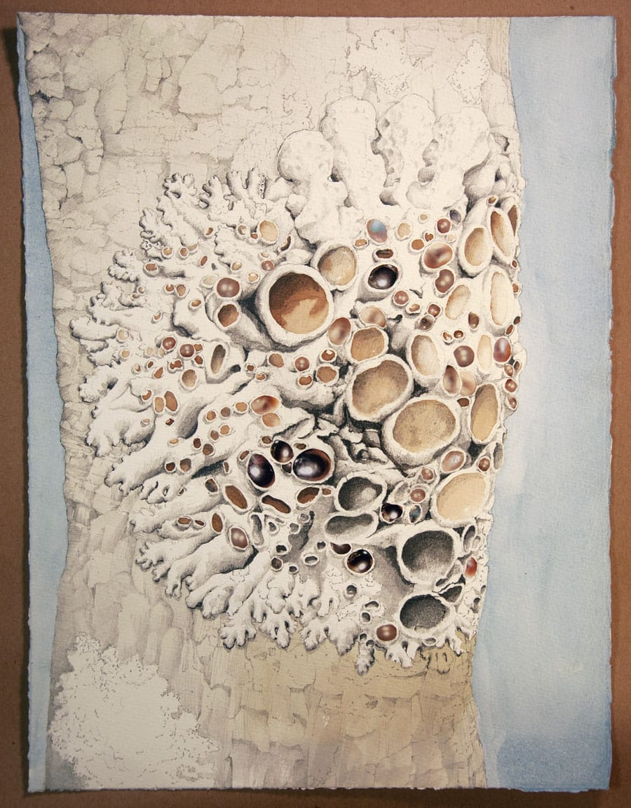

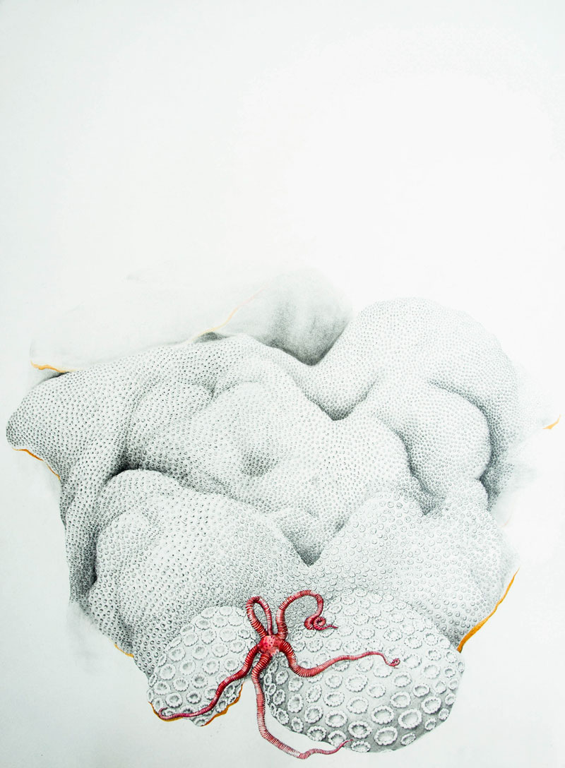
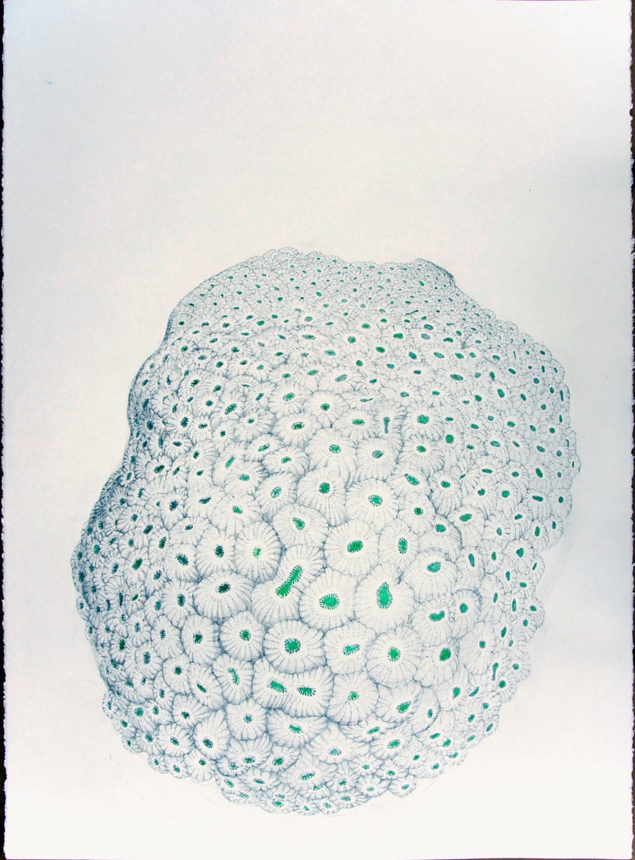
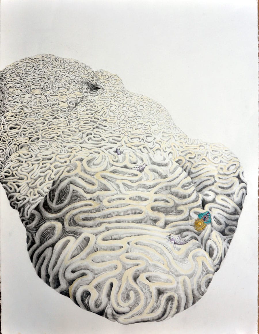
Coral
- Mountainous star (Orbicella faveolota)
-
Sunray lettuce (Helioseris cucculata)
- Great star (Montrastraea)
- Symmetrical brain (Pseudodiploria strigosa)
- Spiny flower (Mussa angulosa)
Between 2019 and 2021, I studied and documented coral reef and their biodiversity from the Flower Garden Banks National Marine Sanctuary, located approximately 100 miles off the Galveston, Texas coast. The resulting drawings were exhibited at the Houston Public Library in Colleen Maynard: Calyxes and Polyps (Celebrating Coral of the Flower Garden Banks National Marine Sanctuary), March-May 2022. This exhibition was funded in part by the City of Houston through Houston Arts Alliance.
![1]()
![2]()
![3]()
![4]()
![5]()